The Cardiovascular system, Heart Muscle, The Heart as a pump
The Cardiovascular System is a closed system, with a one way circuit.
- efficiency
Transports Materials throughout the body:
- Exogenous
- Endogenous
- Waste and heat elimination
- Brain is Oxygen dependent
- Cannot meed oxygen needs by using the anaerobic pathway
- 2 ATP + 1 NADH
- Must use the aerobic pathway - Glycolysis, Citric Acid cycle + Electron Transport System
- 6H2O+ 30 ATP + 6CO2
- Nutrient, Hormone, Immunity transport
- CO2 and metabolic waste and heat removal

How Blood flows:
- Down a pressure Gradient (dP) from high pressure to a low pressure
- Pressure, Volume, Flow and Resistance
- Pressure of a fluid in motion decreases over distance
- Hydrostatic Pressure: = may also be referred to as the hydraulic pressure - as fluid is in motion
- In a system that flows, pressure falls over distance as energy is lost to resistance / friction
- Pressure in Liquid changes without changes in Volume
- Driving pressure - the pressure of the ventricles on the systemic circulation - displaces the blood in a forward direction
- The constriction of the blood vessels also increases blood pressure
- Blood flows from high pressure to low pressure
- Flow depends on or is proportional to the pressure gradient and not the absolute pressures.
- Flow proportional to dP
- Resistance Opposes flow
- Flow is inversely proportional to resistance
- Flow ~ 1/R
- Poiseuille's Law
- Resistance is proportional to the Length, the viscosity of the fluid and inversely proportional to the radius of the tube qubed
- Vasoconstriction has a huge impact on the resistance to blood flow
or
- where:
- ΔP is the pressure drop, L is the length of pipe, μ is the dynamic viscosity, Q is the volumetric flow rate, r is the radius, d is the diameter, π is the mathematical constant
- Velocity depends on the Flow Rate and the Cross-Sectional Area
- Flow rate is not the Velocity of flow
- Flow rate is 5L/min - how much
- Velocity is a measure of how fast blood flows past a point in time
- Velocity of Flow = Flow Rate (Q) / Cross Sectional Area (A)
- v = Q/A
- Velocity is faster in narrow sections and slower in faster sections
- The Mean Arterial Pressure is maintained by the Aorta in diastole. MAP is the primary driving force for blood flow.
- MAP is CO (volume of Blood / minute) x peripheral resistance
- Mean arterial pressure can be determined from:[2]
where:
- CO is cardiac output
- SVR is systemic vascular resistance
- CVP is central venous pressure and usually small enough to be neglected in this formula.
- The mean blood pressure ranges from a high of 93 mm Hg in the Aorta to a low of a few mmHg in the vena cavae


Cardiac Muscle and the Heart
- The heart has its base (up) opposite to the apex (down)
- The lubricating pericardium fluid encases the heart covered by a tough membranous pericardium.
- The heart chambers - left separate from right - but contract in a coordinated fashion - first the atria and then the ventricles.
- The ventricles contract from the bottom up - as blood enters and leaves the ventricles from the top.
- Papillary muscle anchor the chordae tendinae and prevent prolapse of the tricuspid or mitral valves
Cardiac Muscle Cells Contract without innervation
- Contraction is myogenic - originates in the muscle itself - autorhythmic cells or pacemakers
- Myogenic autorhythmic cells do not contract - and are not like other contractile myocardial cells - smaller and contain few contractile fibers
- Contractile myocardial cells - typical striated muscle - with contractile fibers arranged in sarcomeres.
- Intercalated disks are made up of desmosomes to bind the muscle together and gap junctions to facilitate electrical conduction
- Larger T Tubules than those of Skeletal muscle, and they branch inside the ,myocardial cell.
- Sarcoplasmic Reticulum is smaller - depends on extracellular Ca2+ to initiate contraction.
- Mitochondria occupy a 1/3 of the cytoplasmic volume - reflecting the high energy requirement of cardiac cells.
Calcium Entry is a feature of Cardiac Excitation Contraction Coupling
- Calcium induced calcium release
- RyR Channels open in response to Calcium Binding
- Calcium binds to troponin to initiate contraction
- Calcium reenters the Sarcoplasmic Reticulum - with ATP
- Calcium is pumped out using the Sodium Calcium Exchanger (NCX)
- Calcium out for 3 Sodium in down the electrochemical gradient
- Skeletal muscle L-Type Calcium Channels - also called DHP receptors remain Closed but are mechanically linked to the RyR Calcim release channels of the SR.
- Myocardical L-type Calcium Channels open to allow Calcium into the cell. In both muscles, SR calcium channels are associated with RyR Calcium release channels on the SR
- The myocardial cells contract in the presence of extracellular calcium - they do not have large ER stores for contraction. But Skeletal muscles do not require extracellular calcium.
CARDIAC Muscle Contraction can be Graded
- Force is proportionate to the number of cross bridges that are active - the number of cross bridges are determined by the calcium binding to troponin.
- Low calcium - low bridges - small force
- More calcium - more bridges - stronger force
- Tension generated is directly proportional to the initial length of the muscle fibre.
- Cardiac stretch is a function of the amount of blood in the chambers of the heart.
Myocardial Action Potentials Vary
- Myocardial Contractile Cells
- Action potentials are longer because of calcium entry
- Phase 4 - resting membrane potential -90 mV
- Phase 0 - fast double voltage gated Sodium Channels open, depolarising the membrane to +20 mV
- Phase 1 - Potassium leaves through potassium Channels
- Phase 2 - Closing the "fast potassium" channel and Calcium Channels open - causing the plateau
- Phase 3 - rapid repolarisation - calcium channel closes and the potassium permeability increases - "slow potassium" open.
- The calcium entry allows ventricular filling and prevents cardiac tetanus
- Cardiac Muscle prevents tetanus by the prolonged action potential - there is no summation of action potentials like in skeletal muscle as the cardiac muscle is relaxed before the next contraction
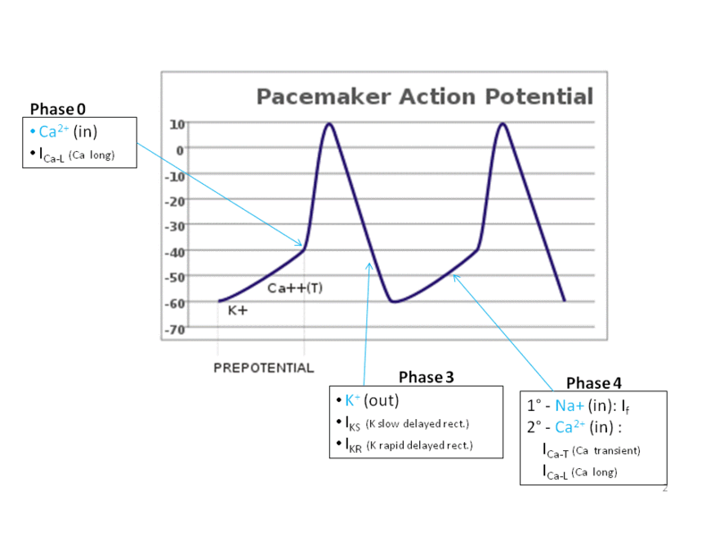
- Myocardial Autorhythmic Cells
- Generation of action potentials spontaneously without nervous system input - UNIQUE
- Never rests or at a constant potential - pacemaker potential - no resting potential -
- -60 mV, If Channels - I is for current and f is for funny - funny current If
- Some Calcium channels then open - calcium then depolarises creating steep depolarisation (Not Sodium!) followed by slow potassium channels (potassium out repolarising the membrane)
- Autonomic Neurotransmitters Modulate Heart Rate
- Sympathetic stimulation from norepinepharine from sympathetic neurons and epinepharine from the adrenal medulla increase the flow of both If and calcium channels. More rapid entry, increase rate of depolarisation, increases rate of action potential, the heart rate increases.
- Catecholamines exert their effect by binding to and activating B1-adrenergic receptors on the autorythmic cells. cAMP - alters the transport properties on the ion channels. In If Channels the cAMP is itself the messenger. When cAMP opens the IF Channels they remain open longer
- The parasympathetic neurotransmitter acetylcholine (ACh) slows the heart rate. ACh activates muscarinic cholinergic receptors that influence Potassium and Calcium channels in the pacemaker cells. Potassium permeability increases and decreases Calcium permeability - hyperpolarising the pacemaker cells. This decreases the rate of depolarisation - as it takes longer to reach threshold - and decreases the rate.

The Heart as a Pump
- Electrical conduction in the Hearth Coordinates Contraction
- SA node - branched intranodal pathway - AV node - Purkinje fibers - AV bundle (bundle of His) - Bundle Branches
- The apex-to-base contraction of the heart allows the heart to to squeeze blood out to the top of the ventricles
- The spiral arrangement of the muscles in the wall of the ventricles aids the ejection of blood from the ventricles - as they pull the apex and the base of the heart closer together, squeezing blood out to the openings at the top of the ventricles.
- Apex node delay promotes ventricular filling.
- Pacemaker set the Heart Rate
- The SA node set the pace of the heartbeat
- The ECG Reflects Electrical Activity
- In lead one the left arm is designated as positive and the right arm as negative
- A single contraction relaxation cycle is known as a CARDIAC CYCLE.
- Waves: deflections above or below the baseline - 3 waves
- P wave: depolarised atria
- QRS Complex: progressive wave of ventricular contraction
- T wave: repolarisation of the ventricles
- Segments: are sections of the baseline between 2 waves and segments
- An ECG is an electrical view of a three dimensional object.
- Questions:
- What is the heart rate?
- Is the rhythm of the heart beat regular or Irregular?
- Are all normal waves present in recognisable form?
- Does a QRS complex follow each P wave, and is the PR segment consistent in Length?

- Atrial and Ventricular diastole
- Completion of ventricular filling
- Atrial systole - see some retrograde flow in the veins as there are no one way valves - observed as a pulse in the jugular vein - head and chest elevated at 30 degrees
- Early ventricular contraction and the first heart sound
- closure of the AV valves - 1st heart sound
- Isovolumic ventricular contraction
- The heart pumps: Ventricular ejection
- Ventricular relaxation - 2nd Heart Sound
- Isovolumic Ventricular Relaxation
- X: Change in Volume
- Y: Pressure


Wiggers Diagram

Stroke Volume is the Volume of Blood pumped per Contraction
- Stroke Volume = EDV- ESV
- Stroke volume = 135ml - 65 ml = 70 ml at rest
- Stroke Volume = Volume of blood before a contraction less the volume of blood after the contraction.
Cardiac Output is a measure of Cardiac Performance
- Cardiac output = heart rate x stroke volume
- Cardiac output = 70 bpm x 70 ml/beat = 5040 ml/min
- Cardiac output is the same for both ventricles - Normally
- If one side of the heart begins to fail (cardiac failure) and is not able to pump efficiently, cardiac output becomes mismatched. Blood pools in the circulation behind the weaker side of the heart.
The Heart Rate is modulated by Autonomic Neurons and Catecholamines:
- The intrinsic heart rate is 90bmp
- Tonic control of the heart rate is dominated by the parasympathetic nerves - slowing the intrinsic heart rate from 90 bpm to 70 bpm.
- Increase the rate by removing parasympathetic activity - cells will return to their intrinsic rate of 90 bpm or activate the sympathetic nervous system with norepinepharine from the nerves or epinepharine via the medulla of the adrenal cortex - act on B1 receptors - speeds up depolarisation and increases heart rate
- Both autonomic branches alter the rate of conduction through the AV node.
- ACh - Parasympathetic Nerves - slows the conduction of action potentials through the AV node and increases AV node delay
- Epinephrine and norepinepharine enhance conduction of action potentials through the AV node and decreases AV delay
Factors that Influence Stroke Volume:
- Stroke Volume is the Volume of Blood pumped per Contraction
- Directly related to the force generated by the cardiac muscle during a contraction
- The length of the muscle fibers at the beginning of the contraction, and
- EDV: The volume of blood at the beginning of the contraction determines the length of the muscle
- Frank Starling Law of the Heart = Length-Tension relationships
- Force is directly related to length of the sarcomere
- As the stretch of the ventricle wall increases so does the stroke volume
- Preload = stretch before contraction
- Starling curve:
- X: Stretch indicated by ventricular EDV
- Y: Force indicated by Stroke Volume
- Stroke volume is proportional to EDV, within physiological limits, the heart will pump blood returned to it
- Stroke Volume and Venous Return
- The EDV is normally determined by venous return.
- Venous Return is determined by
- Skeletal Pump: Contraction or compression of the veins returning blood to the heart -
- esp the lower legs
- Respiratory Pump: Pressure changes in the abdomen and thorax during breathing, and
- Inspiration creates:
- a negative intra-thoracic pressure
- Compression on the abdominal contents during inspiration also increases venous flow
- enhances venous return
- Sympathetic innervation of the veins
- Constricts the veins - squeezing more blood out of them to the heart
- Increasing EDV
- The contractility of the heart
- The intrinsic ability of the cardiac muscle to contract at any given length and
- a function of Calcium interaction with the contractile filaments
- Contractility is Controlled by the Nervous and Endocrine System
- Chemicals that affect contractility are inotropic agents and have an Inotropic effect
- Positive Inotropic Effect: Sympathetic Nervous system - Increase the force of contractility
- Digitalis - used since the 18th Century to treat heart failure - increases contractility by slowing calcium removal from the cytosol (unlike the catacholamines that increase the calcium uptake by the SR). Also decreases the cells ability to remove calcium by the NCX - Sodium Calcium Exchanger
- Toxic to the Sodium Potassium Pump
- Negative Inotropic Effect: Parasympathetic System -
- EDV and Arterial Blood Pressure Determine Afterload
- The combined load of the EDV and the arterial resistance during ventricular contraction is known as the afterload
- Increased afterload is found in:
- hypertension
- decreases aortic compliance
- Increased afterload increases the energy required to generate the Stroke Volume - the force of contraction - increasing the muscles need for oxygen and ATP production. Chronic increase in afterload leads to muscular hypertrophy and increasing cardiac wall thickness -
- measure with ultrasound
- Arterial BP - used as an indirect measure of afterload
- Echocardiography - measure ejection fraction - Stroke volume/EDV
- 70ml/135ml or 52% at rest and 74% in exercise

Cardiac muscle cells has intercalated disks between cardiac muscle cells, which is different from skeletal muscle. These discs have very low electrical resistance and allow action potential to travel easily between cardiac muscle
The cardiac muscle is a syncytium of many heart muscle cells in which the action potential spreads rapidly from cell to cell.
The atrioventricular (AV) bundle conducts impulses from the atria to the ventricles. This is an exclusive pathway as the atrial and ventricular syncythium are isolated from one another by fibrous tissue.
Action Potentials in cardiac Muscle
The resting membrane potential of cardiac muscle is about -85 to -95 mV and the action potential is 105 mV. The membranes depolarise for 0.2 seconds in the atria and 0.3 seconds in the ventricles.
Slow entry of Sodium and Calcium ions into the cardiac Muscle is one of the causes of the action potential plateau. In cardiac muscle there are also fast sodium channels that open at the initiation of the action potential, but cardiac muscle has a unique slow calcium channel or calcium sodium channel. Through the slow channels the calcium and the sodium ions flow into the cell after the initial spike of the action potential, and they maintain the plateau. Calcium enters the cells through this channel and promotes muscle contraction.
Another cause for the plateau of the action potential is the decrease in the permeability of cardiac muscle cells to potassium ions. This decrease in potassium permeability also prevents the return of the membrane potential. When the calcium channel closes after 0.2 to 0.3 seconds the potassium permeability increases rapidly and the membrane potential returns to its resting level.
Diffusion of calcium into the myofibrils promotes muscle contraction. The action potential that spreads into the each cardiac muscle fiber along the transverse T Tubules and this causes longitudinal sarcoplasmic tubules to release calcium ions into the sarcoplasmic reticulum. This stimulates muscle contraction - like skeletal muscle.
The T Tubules of cardiac muscle have 25 x greater storage of calcium then skeletal muscle and this is released during the action potential. At the end of the action potential the calcium influx is stopped, the calcium is pumped back into the sarcoplasmic reticulum and T Tubules, and contraction ends.

Cardiac action potentials
Main articles: Cardiac action potential, Electrical conduction system of the heart, Cardiac pacemaker, and Arrhythmia

The cardiac action potential differs from the neuronal action potential by having an extended plateau, in which the membrane is held at a high voltage for a few hundred milliseconds prior to being repolarized by the potassium current as usual.[93] This plateau is due to the action of slowercalcium channels opening and holding the membrane voltage near their equilibrium potential even after the sodium channels have inactivated.
The cardiac action potential plays an important role in coordinating the contraction of the heart.[93] The cardiac cells of the sinoatrial nodeprovide the pacemaker potential that synchronizes the heart. The action potentials of those cells propagate to and through the atrioventricular node (AV node), which is normally the only conduction pathway between the atria and theventricles. Action potentials from the AV node travel through the bundle of Hisand thence to the Purkinje fibers.[note 2] Conversely, anomalies in the cardiac action potential—whether due to a congenital mutation or injury—can lead to human pathologies, especially arrhythmias.[93] Several anti-arrhythmia drugs act on the cardiac action potential, such as quinidine, lidocaine, beta blockers, andverapamil.[94]
The Cardiac Cycle

The top three curves show the aortic pressure, left ventricular pressure, and left atrial pressure. The curves below these are changes in ventricular volume, the electrocardiogram, and the phonocardiogram, a recording of heart sounds.
The atria function as primer pumps for the ventricle. 75% of the ventricular filling occur during diastole before the contraction of the atria, which causes the remaining 25% ventricular filling. In AF or SVT there is very little dyspnoea.
The aterial pressure waves:
- a wave: caused by atrual contraction,
- c-wave: caused by ventricular contraction - AV bulging
- v-wave: caused by the in-filling of the atria by venous return.
The ventricles fill with blood during diastole.
- During systole, the AV velves are closed, and the atria fill with blood.
- At the beginning of diastole, when ventricular pressure decreases below the atria, the AV valves open.
- The higher the pressure in the atria pushes blood into the ventricles during diastole.
- The period of rapid filling of the ventricles occurs during the first 1/3 of diastole and provides most ventricular filling.
- Atrial contraction occurs during the last 1/3 of diastole and contributes 25% of filling of the ventricle.
Outflow of blood from the ventricles occur during systole.
- At the beginning of systole, ventricular contraction occurs, the AV valve closes, and the pressure builds up in the ventricle. No outflow of blood occurs in the first 0.2 to 0.3 seconds - persiod of isovolumic contraction,
- When LV pressure > aortic pressure of 80mmHg, the RV pressure > Pulmonary pressure of 8mmHg, the aortic and pulmonary valves open. Ventricular outflow occurs - period of ejection.
- Most ejection occurs in the first part of this period of ejection.
- The last part of systole - period of isovolumic relaxation - and is caused by ventricular relaxation, which causes the ventricular pressure to fall below the aortic and pulmonary artery pressures. Thus, the semiluner valves close.
The fraction of end-diastolic volume that is ejected is called the ejection fraction.
- At the end of diastole the volume of each ventricle is 110 to 120 ml; this is the end diastolic volume.
- The stroke volume, which has a value of 70ml is the amount of blood that is ejected with each beat.
- The end-systolic volume is remaining volume in the ventricle at the end of systole - 40 to 50ml.
- Ejection fraction = Strove Volume (70 ml) / end-diastolic volume (110 to 120 ml) = 60%.
- By increasing the end-diastolic volume and decreasing the end systolic volume, the stroke volume can be doubled.
Ventricular ejection increases pressure in the aorta to 120mmHg (systolic pressure).

Work output of the Heart.
The STROKE WORK OUTPUT of the ventricles is the output of energy by the heart during each heart beat.


 When Venous Return of blood increases, the heart muscle stretches more, which makes it pump with greater force of contraction. The Frank-Sterling mechanism can be stated in a different way - within physiological limits, theheart pumps all the blood that comes to it without allowing the excess accumulation of blood in the veins.


The STROKE WORK OUTPUT of the ventricles is the output of energy by the heart during each heart beat.
- The volume-pressure work of the heart is the work done to increase the blood pressure of the blood.; in the left heart = stroke volume (70ml) x (left ventricular mean ejection pressure and the left ventricular mean input pressure). Right heart volume work pressure is 1/6 of the L heart.
- The kinetic energy supplied to the blood: 1/2MV2. The kinetic energy is only 1%, but in Aortic stenosis the kinetic energy may increase to 50% of the total work output.
The volume-pressure diagram of the left ventricle determines the cardiac work output:
intraventricular pressure as a function of the left ventricular volume
The different phases of the cardiac cycle are:
Phase I: Period of filling during which the ventricular volume increases from end-systolic volume to the end-diastolic volume, or from 45ml to 115ml, an increase of 70ml.
Phase II: Period of isovolumic contraction during which the volume of the ventricle remains at the end-diastolic volume but the intraventricular pressure increases to aortic diastolic pressure, or 80mmHg.
Phase III: Period of ejection during which the systolic pressure increases further because of additional ventricular contraction, and ventricular volume decreases by 70ml, which is the stroke volume.
Phase IV: Period of isotonic relaxation during which the ventricular volume remains consistent at 45ml but the intravascular pressure decreases to its diastolic level.
The area inside the pressure volume curve represents the volume-pressure work (or external work output) if the ventricle during each cardiac cycle. Cardiac work is affected by the PRELOAD AND THE AFTERLOAD on the heart. Preload is the end-diastolic pressure, and the after load is the pressure in the artery exiting the ventricle (aorta or pulmonary artery).
In cardiovascular physiology, end-diastolic volume (EDV) is the volume of blood in a ventricle at the end of filling (diastole). Because greater EDVs cause greater distention of the ventricle, EDV is often used synonymously with preload, which refers to the length of the sarcomeres in cardiac muscle prior to contraction (systole). An increase in EDV increases the preload on the heart and, through the Frank-Starling mechanism of the heart, increases the amount of blood ejected from the ventricle during systole (stroke volume).
Oxygen consumption by the heart is dependent on the cardiac work. Cardiac oxygen consumption mainly depends on the volume-pressure type of work. Oxygen consumption is proportionate to the tension of the heart x the time the tension is maintained. WALL Pressure in the heart is proportional to the pressure x the diameter of the wall. Ventricular wall tension, therefore, increases at high systolic pressures or when the heart is dilated.
Regulation of Heart Pumping
The Frank-Starling mechanism intrinsically regulates cardiac pumping ability.
 When Venous Return of blood increases, the heart muscle stretches more, which makes it pump with greater force of contraction. The Frank-Sterling mechanism can be stated in a different way - within physiological limits, theheart pumps all the blood that comes to it without allowing the excess accumulation of blood in the veins.
 The extra stretch of cardiac muscle during increased venous return, within limits, causes the actin and myosin filaments to interdigitate at a more optimal length for force generation. In addition more stretch of the right atrial wall causes a reflex increase in the heart rate of 10 to 20%, which helps the heart pump more blood.
Physiology
As the heart fills with more blood than usual, the force of cardiac muscular contractions increases.[2] This is a result of an increase in the load experienced by each muscle fibre due to the extra blood load entering the heart. The stretching of the muscle fibres augments cardiac muscle contraction by increasing the affinity oftroponin C for calcium, causing a greater number of actin-myosin cross-bridges to form within the muscle fibres. The force that any single cardiac muscle fiber generates is proportional to the initial sarcomere length (known as preload), and the stretch on the individual fibers is related to the End Diastolic Volume of the left and right ventricles.
In the human heart, maximal force is generated with an initial sarcomere length of 2.2 micrometers, a length which is rarely exceeded in the normal heart. Initial lengths larger or smaller than this optimal value will decrease the force the muscle can achieve. For larger sarcomere lengths, this is the result of less overlap of the thin and thick filaments; for smaller sarcomere lengths, the cause is the decreased sensitivity for calcium by the myofilaments.
Cardiac and Vascular Function Curves
1. Mean Systemic Pressure Changes: The mean systemic pressure is affected by blood volume as well as venous compliance. Changes in the mean systemic pressure will shift the vascular function curve left or right.
2. Inotropic Changes : Contractility is determined by various autonomic mechanisms and certain drugs (such as digitalis). Inotropic changes will alter the slope of the cardiac curve up or down (as discussed above).
Positive inotropic agents, such as digoxin, will increase contractility and therefore increase the cardiac output (as shown above). This new equilibrium point now reflects an increased cardiac output and a lower right atrial pressure (more blood is now being ejected from the heart with each beat).
Negative inotropic agents have the opposite effect, decreasing contractility and cardiac output, and increasing right atrial pressure (not shown).
3. Total Peripheral Resistance Changes : TPR is determined by the resistance of the arterioles. Changes in TPR will change the slope of both the cardiac function curve and the venous return curve.
- An increase in TPR (shown above) will cause blood to be retained on the arterial side of circulation and will increase the aortic pressure against which the heart must pump. This will act to shift both slopes downward. As a result of this simultaneous change, both the cardiac output and the venous return are decreased, however the right atrial pressure remains the same .
A decrease in TPR (not shown) will allow more blood to flow to the venous side of circulation and will lower the aortic pressure against which the heart must pump. This will shift both slopes upward. Both cardiac output and venous return will be simultaneously increased; again, right atrial pressure will remain the same .
The autonomic nervous system affects cardiac pumping. Sympathetic stimulation increases the heart rate from 72bpm to 180bpm and dramatically increases the force of cardiac contraction. Sympathetic stimulation can increase cardiac output 2 to 3 fold.
Parasympathetic stimulation mainly decreases atrial heart rate dramatically but only slightly decreases the force of ventricular contraction. The combine effect is a decrease in cardiac output by 50%.
Cardiac contractility is affected by several factors.
Electrolyte concentrations - excessive K+ causes the heart to become flaccid and reduces heart rate decreasing contractility.
Excessive calcium causes the heart rate to go into spastic contraction. Decrease in calcium make the heart flaccid.
Rate of change of cardiac contractility = dP/dt, and is effected by the Preload and the afterload. Another index is the (dP/dt)/P
http://www.cytokinetics.com/cardiovascular_program

The cardiovascular system refers to the heart, blood vessels and the blood. Blood contains oxygen and other nutrients which your body needs to survive. The body takes these essential nutrients from the blood. Thanks for explaining in details.
ReplyDeletebuy tramadol online | buy enclomiphene | dapoxetine for sale | sildenafil citrate online
Great post.
ReplyDeletehttps://snipplr.com/users/dorianperry
Good one! Thanks for sharing. By the way What's the benifit of investing in funds over the individual stocks and bonds?
ReplyDeleteSensex
Sensitive Index
BSE Sensex
Thank you so much for sharing this information. NOVAREL® (chorionic gonadotropin for injection, USP) is a prescription medicine that contains a hormone to help stimulate healthy ovaries to make eggs. NOVAREL is used for women who need medical help to get pregnant. Your doctor may prescribe more than one medicine as part of a pregnancy plan. Buy NOVAREL from onlinegenericmedicine.com (Online Pharmacy), they provide all types of generic medicine, best in class products and best deals. Buy medicines and get the generic medicines delivered in the USA, UK & Australia. Click here to Get more info about these medicines.
ReplyDeleteGreat post.
ReplyDeletehttps://site-stats.org/7dmc.ae/
Hi Dear,
ReplyDeleteI read your post and fund it very informative as well helpful. Thanks for sharing this informational blog post. For more information click this hyper link Phases of the Cardiac Cycle
Nice Informative Blog having nice sharing..
ReplyDeletefitness
Great post.
ReplyDeletehttps://sketchfab.com/Vmeals
Great post.
ReplyDeletehttp://www.genina.com/user/profile/1670923.page